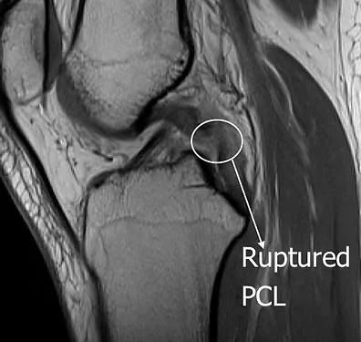
MRI scan showing a PCL rupture
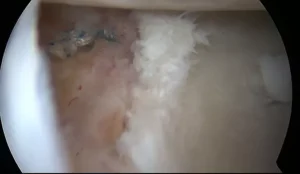
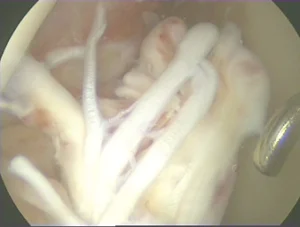
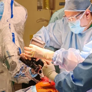
PCL reconstruction is also performed through arthroscopic surgery similar to ACL reconstruction. In arthroscopic PCL reconstruction, small key-hole sized incisions are used and the PCL is reconstructed under direct view with an arthroscopic camera. Like the ACL, the PCL which is ruptured cannot be simply stitched back together. It has to be reconstructed, meaning replaced using a tendon graft. This tendon graft is usually taken from the patient’s own tendons. Once the tendon graft has been placed in the original position of the natural PCL, the graft is secured to the bones using special screws which are bio-absorbable, meaning that they will disappear after a few years. By then, the PCL graft would have grown secured into the bone and the screws no longer required. During the same surgery, other ruptured ligaments like the PLC or LCL can also be reconstructed together with the PCL. Reconstruction of the PCL and other complex ligaments like the LCL or PLC need to be done early in order to obtain the best outcomes.

Arthroscopic camera showing a reconstructed PCL
In cases where a PCL and PLC / LCL injury is chronic or neglected (injury period exceeding 6 weeks of), ligament reconstruction by itself is not sufficient to restore the knee back to normal. Knee osteotomy surgery (high tibial osteotomy) is required to treat chronic cases. In many cases, high tibial osteotomy alone is sufficient to restore stability of the knee so that the patient does not need an additional knee ligament reconstruction. Now, with the use of computer-guided surgery (CAS), high tibial osteotomy can be performed with better accuracy and more consistent results.


